Virtual Reality Assisted Imaging in Complex Congenital Heart Surgery: Scope and Prospects
April 24, 2024 | Contributed by Dr Balaji Srimurugan

The role of three-dimensional imaging has made its way into every sphere of life, be it in entertainment / education or a myriad of other uses. From wearable 3D glasses to 3D gaming to 3D software in defence intelligence, it’s presence is everywhere and is here to stay. Virtual Reality is a simulated experience that employs pose tracking and 3D near eye displays to give the user an immersive feel of the virtual world. Virtual reality has made it big in the field of entertainment, education and business. A person using virtual reality equipment is able to look around the artificial environment and interact with the virtual features and items.
The evolution of congenital heart disease and children heart treatment in particular has been made possible because it allowed a healthy marriage of medical knowledge with engineering technology. The role of medical imaging in the accurate diagnosis of congenital heart disease to precisely guide its treatment forms an integral part in the management of congenital heart disease.
The Role of Virtual Reality
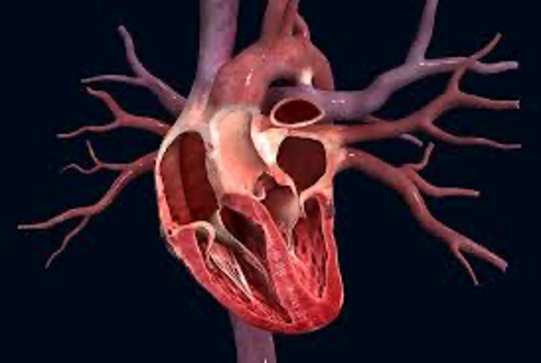
However, the two-dimensional imaging that the Echocardiography and the computerised tomography offer, has it’s own limitations particularly in the assessment of congenital heart conditions that are rare and more complex than the more commonly seen defects. Virtual reality has found its way in redefining the medical imaging of complex congenital heart defects. A few institutes that perform high end congenital heart surgery for complex CHDs have adopted this technology to a great extent. VR technology in the field of children heart treatment has a special role to play in the management of very complex heart defects which are not amenable to be studied by the conventional imaging methods such as echocardiography and computerised tomography.
 The scope of Virtual Reality in treating heart defect is tremendous in the future
The specific applications of Virtual reality assisted imaging modalities in congenital heart surgery can be summarised as follows:
- Those complex heart defects that have been ruled out as inoperable or not suitable for correction can be better studied in depth with the assistance of VR assisted imaging. This may pave way for the revision of the initial decision of inoperability.
- When complex lesions are studied pre operatively by the surgeons with the help of VR assisted imaging, they are made to have a more thorough understanding of the cardiac lesion which enables them to have more surgical precision on the operating table, which in turn translates into shorter operating time and better congenital heart defect treatment for children.
- The VR assisted imaging breaks the barrier of the learning curve that is essential to understand the 2D imaging. Just with the knowledge of the basic anatomy, a fresher and an expert can both interpret the image with equal ease and confidence.
- The application of VR and its scope in the future is tremendous and it can be a great tool for helping those patients with complex heart defects who have been written off as inoperable or impossible. The immersive understanding of the cardiac anatomy greatly serves to improve the confidence of the surgeon in dealing with even the rare and complex anatomies.
- Extended use of the VR imaging can be applied for international collaboration with physicians and surgeons when they conference together in discussing heart conditions. Each expert can meet together as their “avatar” in the virtual space and enter into the immersive experience of “understanding the heart” with its most complex challenges.
To summarise, the use of the technology in the field of medical science and congenital heart defect treatment for children is going to keep changing the paradigm of clinical practice. With Artificial Intelligence making its way into the foray of medicine, it is upto the practicing clinician to apply the technology in the best way possible such that it translates into a patient care.
Dr Balaji Srimurugan,
Assistant Professor,
Division of Pediatric CVTS, AMRITA Kochi

